
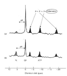
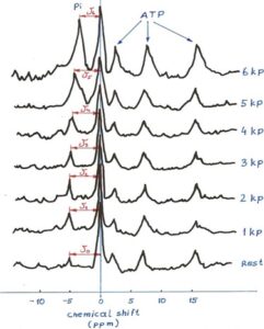
The only possibility for measurement muscle metabolism was possible by using Magnetic Resonance Spectroscopy (MRS) with collaboration with Department of Magnetic Resonance at Jožef Stefan Institute. The MRS technology analysis typical frequency spectrum of Phosphorus nucleus spinning return to its normal spin after its short interval of disturbing with specific frequency of radio waves. The principle of measuring represented graph below.
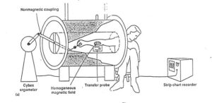
Because Phosphorus (P) atoms are important part of typical high energetic compounds: Adenosine Tri Phosphate (ATP), Creatine Phosphate (CrP) and free Phosphate (Pi), the measurements of non-invasive metabolism of muscle metabolism by using these compounds is possible. The typical P spectrum of muscle enable observation of metabolism of exercising muscle during contractions, however only if muscle under observation is not moving. Our special interest is observing muscle pH, which by using specific ppm shift (δ)in CrP and Pi enable calculation of muscle pH.
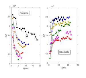
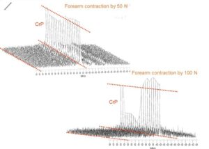
Using arbitrary values of ATP, CrP, and Pi it is possible to observe also the time course of changes of these substances during exercise and therefore time course (dynamics) of changes. A 3-D presentation of these changes during exercise provide next step in ascertain of muscle metabolism of high energy phosphates. From pre exercise resting level on the left part of both diagrams, muscle CrP decrease and remained steady during futher unchanges force of 50 N (upper figure). In contrary, the contraction force of 100 N decreased CrP more than twice in a manner where CrP steady state was no longer possible. During recovery CrP increased and reached values typically higher than during pre-exercise resting interval (supercompensation).